Neural Quantification and Imaging Core
The primary objectives of the Neural Quantification & Imaging Core within the Center for Pediatric Brain Health are to provide state-of-the-art instrumentation and analysis support to the research project leaders (RPLs) and other neuroimaging scientists within the Institute for Human Neuroscience and beyond. The Core is directed by Dr. Tony Wilson, who has over 20 years of experience in human neuroimaging, with expertise stretching across multiple neuroimaging modalities, data analytic approaches, and participant populations. The Neural Quantification & Imaging Core provides researchers with the below resources:
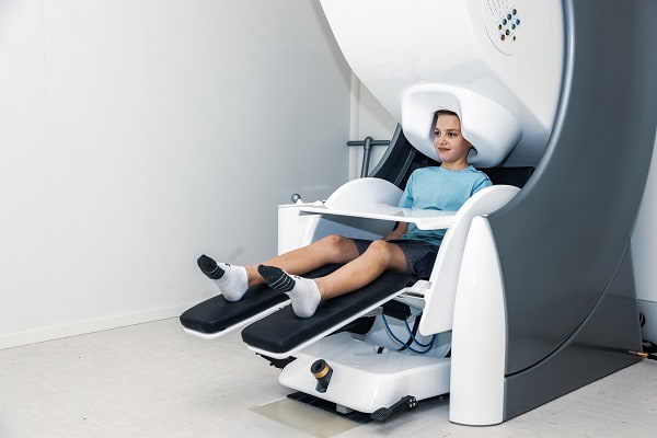
Magnetoencephalography (MEG)
The brand-new Institute for Human Neuroscience facility houses two state of the art MEGIN Triux Neo MEG machines and an expansive collection of magnetically-silent stimulus presentation equipment for cognitive neuroscience type experiments. The instruments are housed within rooms equipped with active shielding and includes the latest technology for head motion correction. The facility was just opened in early 2021.
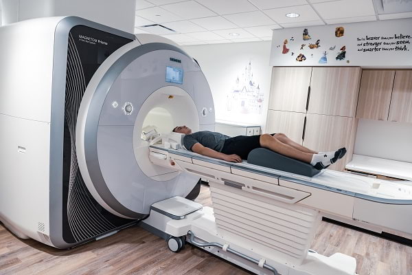
Magnetic Resonance Imaging (MRI)
The Center for Pediatric Brain Health also houses a brand new research-dedicated 3-Tesla Siemens Prisma MRI scanner, equipped with 20-, 32-, and 64-channel head coils, as well as other coils for cardiac, spinal, and other types of imaging. The instrument is configured with multiband imaging capabilities and many experimental, research-only, and commercial sequences, including numerous MRI sequences for advanced diffusion, spectroscopy, structural, and functional brain imaging. The system has the [204×64] XR 80/200 gradients (the most powerful commercially available gradients). The Prisma MRI Suite is also equipped with the necessary peripherals to present experimental stimuli and acquire behavioral responses, including a 32" in-room LCD monitor for presenting stimuli and multiple ergonomic subject response devices.
Brain Stimulation
The Center for Pediatric Brain Health is also equipped with state-of-the-art electrical brain stimulation equipment, including three Soterix Medical systems. The suite includes a standard two-pad tDCS system, a two-pad transcranial electrical stimulation (tES) system, a five-lead multipolar high-definition tDCS system (HD-tDCS), and a five-lead alternating-current stimulation (tACS) system. All of the systems are equipped with settings for sham-stimulation, which allows investigators to use “placebo-controlled" experimental designs. In addition to the stimulators, there is a Polhemus digitizer for coregistering the stimulation sponges or metal electrodes to neuroanatomical images. Users also have access to advanced software for finite-element modeling (FEM) of current flow using the participant's individual anatomy.
Data Processing
The Center for Pediatric Brain Health includes a high-performance computing space. This space currently includes over 50 high-performance workstations for data processing, a 36 terabyte (RAID5) storage array for MEG and MRI data, and a video conferencing system for virtual meetings. Each computer has Matlab and other important software for euroimaging and statistical analyses, including many packages such as SPM, FSL, AFNI, FreeSurfer, CONN, R, and other leading toolboxes. Many of the computers are also equipped with the Brain Electrical Source Analysis (BESA) software, SPSS, and current-distribution modeling software. The open concept space encourages collaborative programming (e.g., algorithm development) and data processing efforts among students and faculty.